
Archivo:Fundus of patient with retinitis pigmentosa, mid stage.jpg
Tamaño de esta previsualización: 699 × 599 píxeles. Otras resoluciones: 280 × 240 píxeles · 560 × 480 píxeles · 871 × 747 píxeles.
Ver la imagen en su resolución original (871 × 747 píxeles; tamaño de archivo: 107 kB; tipo MIME: image/jpeg)
| Descripción |
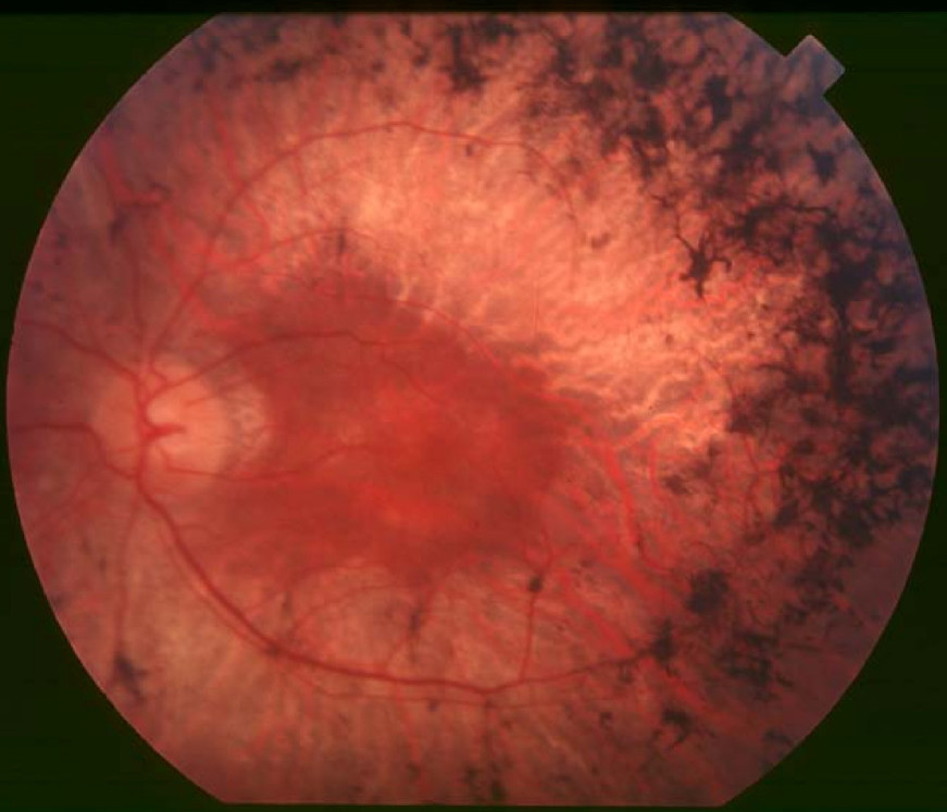
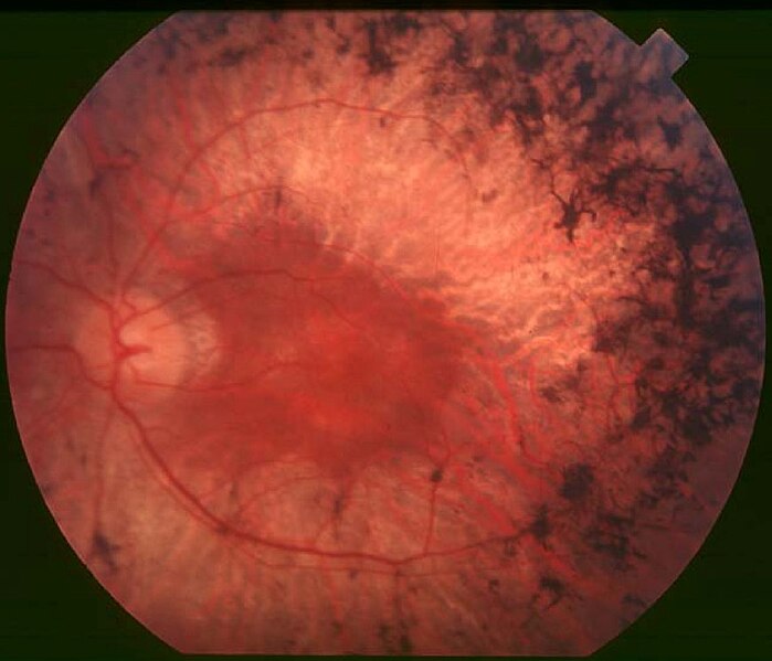
English: Figure 2. Fundus of patient with retinitis pigmentosa, mid stage (Bone spicule-shaped pigment deposits are present in the mid periphery along with retinal atrophy, while the macula is preserved although with a peripheral ring of depigmentation. Retinal vessels are attenuated.) Hamel Orphanet Journal of Rare Diseases 2006 1:40 doi:10.1186/1750-1172-1-40 |
| Fecha | |
| Fuente | Retinitis pigmentosa by Christian Hamel |
| Autor | Christian Hamel |
| Permiso (Reutilización de este archivo) |
© 2006 Hamel; licensee BioMed Central Ltd. This is an Open Access article distributed under the terms of the Creative Commons Attribution License (https://creativecommons.org/licenses/by/2.0), which permits unrestricted use, distribution, and reproduction in any medium, provided the original work is properly cited. |
Este archivo está disponible bajo la licencia Creative Commons Atribución 2.0 Genérica.
- Eres libre:
- de compartir – de copiar, distribuir y transmitir el trabajo
- de remezclar – de adaptar el trabajo
- Bajo las siguientes condiciones:
- atribución – Debes otorgar el crédito correspondiente, proporcionar un enlace a la licencia e indicar si realizaste algún cambio. Puedes hacerlo de cualquier manera razonable pero no de manera que sugiera que el licenciante te respalda a ti o al uso que hagas del trabajo.
Historial del archivo
Haz clic sobre una fecha y hora para ver el archivo tal como apareció en ese momento.
| Fecha y hora | Miniatura | Dimensiones | Usuario | Comentario | |
|---|---|---|---|---|---|
| actual | 10:17 2 dic 2017 | | 871 × 747 (107 kB) | Doc James | Cropped 27 % horizontally and 7 % vertically using CropTool with precise mode. |
| 13:52 22 sep 2009 |  | 1200 × 799 (126 kB) | CopperKettle | {{Information |Description={{en|1=Figure 2. Fundus of patient with retinitis pigmentosa, mid stage (Bone spicule-shaped pigment deposits are present in the mid periphery along with retinal atrophy, while the macula is preserved although with a peripheral |
Usos del archivo
Las siguientes páginas usan este archivo:
Uso global del archivo
Las wikis siguientes utilizan este archivo:
- Uso en ar.wikipedia.org
- Uso en bs.wikipedia.org
- Uso en ca.wikipedia.org
- Uso en da.wikipedia.org
- Uso en en.wikipedia.org
- Uso en en.wikiversity.org
- Uso en eu.wikipedia.org
- Uso en fa.wikipedia.org
- Uso en fi.wikipedia.org
- Uso en fr.wikipedia.org
- Uso en he.wikipedia.org
- Uso en hy.wikipedia.org
- Uso en it.wikipedia.org
- Uso en ko.wikipedia.org
- Uso en la.wikipedia.org
- Uso en or.wikipedia.org
- Uso en outreach.wikimedia.org
- Uso en pl.wikipedia.org
- Uso en pt.wikipedia.org
- Uso en ru.wikipedia.org
- Uso en sl.wikipedia.org
- Uso en sr.wikipedia.org
- Uso en sv.wikipedia.org
- Uso en th.wikipedia.org
- Uso en tr.wikipedia.org
- Uso en tt.wikipedia.org
- Uso en uk.wikipedia.org
- Uso en vi.wikipedia.org
- Uso en www.wikidata.org